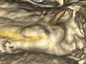
Exposing the facial nerve is one of the most delicate tasks in mastoidectomy. In this step, the bone is removed until facial nerve and chorda tympani become visible, leaving only a thin layer of bone over the nerves.
The left image shows a surgical site in the VOXEL-MAN Tempo surgery simulator, after this step has been performed on a left ear. The course of the facial nerve and the short process of the incus are clearly visible.
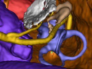
The right image shows exactly the same view, but without the bone. Various structures at risk integrated into the virtual 3D anatomy model of the temporal bone become visible, such as the sigmoid sinus, facial nerve, chorda tympani, tympanic membrane, auditory ossicles, cochlea, semicircular canals and dura mater. Besides their visual and haptic rendering, these model components also allow to detect possible injuries during a virtual surgery.

